
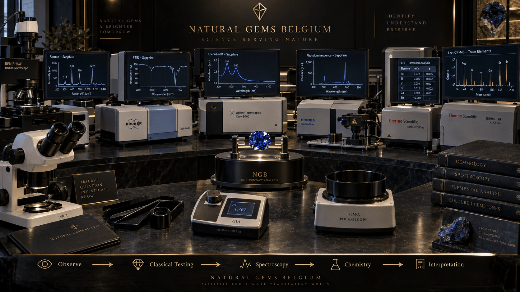
Table of Contents
From Refractive Index to Atomic Fingerprints
A gemstone arrives at a laboratory.
Under the microscope it appears natural.
Its refractive index is correct.
Its specific gravity is correct.
Its optical character is correct.
Its spectrum is broadly compatible with the suspected species.
So the identification is finished.
Or is it?
Perhaps the client also wants to know:
Has this sapphire been heated?
Is beryllium diffusion present?
Is this emerald natural or hydrothermal synthetic?
What is filling its fractures?
What causes this gemstone’s colour?
Does the trace-element chemistry support a particular geographic origin?
Suddenly the familiar instruments of classical gemmology may no longer provide enough information.
The stone has not changed.
The question has.
This is where advanced gemstone laboratory testing begins.
Modern gemmology combines two complementary worlds:
classical observation of physical and optical properties
and
instrumental analysis of molecules, lattice defects, absorption and chemical composition.
Neither replaces the other.
Together they allow gemmologists to investigate a gemstone from its visible inclusions down toward its atomic-scale structure.
PART I — CLASSICAL GEMMOLOGY REMAINS THE FOUNDATION
1. What Can Classical Instruments Tell Us?
A traditional gemmological laboratory may use:
- 10× loupe;
- gemmological microscope;
- refractometer;
- polariscope;
- dichroscope;
- handheld spectroscope;
- ultraviolet lamps;
- hydrostatic weighing.
These instruments remain extraordinarily powerful.
They can reveal:
- refractive index;
- birefringence;
- optical character;
- pleochroism;
- specific gravity;
- fluorescence;
- absorption features;
- inclusions;
- growth structures;
- colour zoning;
- many treatment clues.
For a large proportion of gemstones, this evidence can establish a reliable identification.
Advanced instrumentation should therefore not be imagined as replacing classical gemmology.
It extends it.
2. Why Does Classical Testing Sometimes Reach a Limit?
Consider natural and synthetic sapphire.
Both are corundum.
Both can produce essentially the same:
- RI;
- birefringence;
- SG;
- hardness;
- optical character.
Or consider two natural sapphires from different geological deposits.
Both remain:
Al₂O₃.
Their standard gemmological constants may overlap almost completely.
Yet tiny differences in:
- trace elements;
- lattice defects;
- inclusions;
- growth chemistry
may preserve information about their histories.
Those differences may occur at concentrations far below what the human eye—or a handheld spectroscope—can resolve.
Advanced laboratory instruments are designed to interrogate precisely those differences.
PART II — THINK IN QUESTIONS, NOT MACHINES
3. There Is No Universal “Gemstone Scanner”
A common misconception is that a modern laboratory places a gemstone into one machine that automatically returns:
Species: Sapphire
Origin: Sri Lanka
Treatment: Unheated
Natural: Yes
Real gemmology does not work that way.
Different instruments measure different physical phenomena.
A laboratory therefore chooses methods according to the question.
For example:
What mineral is this?
Raman spectroscopy may help.
What molecular bonds or impurities are present?
FTIR may help.
What causes the colour?
UV-Vis-NIR may help.
Which optical defects exist in the crystal lattice?
Photoluminescence may help.
Which chemical elements are present?
XRF may help.
How much of a trace element is present at very low concentration?
LA-ICP-MS may help.
The result emerges from:
combined evidence + reference data + expert interpretation.
PART III — RAMAN SPECTROSCOPY
4. Asking a Material to Identify Its Structure
Raman spectroscopy is one of the most elegant identification techniques in modern mineralogy and gemmology.
A laser illuminates the sample.
Most photons interact with the material without changing energy.
A very small fraction exchange energy with vibrational modes in the material.
This produces:
Raman scattering.
The resulting spectrum contains peaks characteristic of the material’s atomic structure.
In practical terms, many minerals possess a recognisable:
Raman fingerprint.
5. What Can Raman Identify?
Raman spectroscopy can be extremely useful for identifying:
- gemstones;
- unusual minerals;
- inclusions inside gemstones;
- some treatment materials;
- unknown crystalline phases.
Suppose the microscope reveals a tiny crystal inside an emerald.
Its shape suggests zircon.
Visual morphology alone may not prove that conclusion.
A Raman microscope can focus on the inclusion and compare its spectrum with known reference spectra.
The inclusion can sometimes be identified without extracting it from the gemstone.
That is an enormous analytical advantage.
6. Host and Inclusion Can Both Be Investigated
Raman analysis is particularly powerful when the unknown feature lies:
inside another gemstone.
GIA researchers have used Raman spectroscopy to identify mineral and fluid components inside gemstones.
This turns the microscope from a purely visual instrument into a targeted analytical platform.
Instead of saying:
“This looks like zircon.”
the laboratory may be able to say:
“Its Raman spectrum is consistent with zircon.”
7. Raman Is Not Perfect
Fluorescence can interfere with Raman measurements.
Some materials produce strong luminescence under the laser wavelength, overwhelming the weaker Raman signal.
Laboratories can sometimes solve this by:
- changing laser wavelength;
- adjusting acquisition conditions;
- using another analytical method.
The lesson is already becoming clear:
advanced instruments also have limitations.
PART IV — FTIR SPECTROSCOPY
8. Listening to Atomic Bonds Vibrate
FTIR stands for:
Fourier-Transform Infrared Spectroscopy.
Instead of examining visible colour, FTIR measures how a material absorbs:
infrared radiation.
Infrared energy interacts strongly with vibrations of atomic bonds.
Different bonds absorb characteristic ranges of infrared energy.
The resulting spectrum can therefore reveal information about:
- molecular groups;
- impurities;
- structural defects;
- foreign treatment substances.
9. Why FTIR Became So Important
GIA has used FTIR for gemstone analysis since:
1986.
Today it is one of the most important analytical techniques in advanced gemmology.
Applications include:
- diamond classification;
- natural versus laboratory-grown separation;
- treatment detection in corundum;
- emerald investigations;
- polymer detection in jadeite and turquoise;
- fracture-filling analysis.
The technique is generally nondestructive and can provide substantial information rapidly.
10. Diamond Type
Diamond provides a particularly powerful example.
Nitrogen and boron impurities produce characteristic infrared absorptions.
FTIR allows diamonds to be classified into types such as:
Type Ia
Type Ib
Type IIa
Type IIb
Diamond type alone does not always provide a final natural-versus-laboratory-grown identification.
But it can radically narrow the problem.
For example, GIA notes that nearly all colourless laboratory-grown diamonds are type IIa, while fewer than 2% of natural diamonds belong to that category.
That means:
Type IIa ≠ synthetic
but it can signal that additional testing is especially important.
11. FTIR and Emerald
Natural and hydrothermal synthetic emerald can present a difficult problem because both crystallised in environments involving hot fluids.
Their infrared spectra can consequently share features.
However, FTIR can reveal specific absorptions related to structural components and growth chemistry.
GIA documents chlorine-related infrared peaks in commonly encountered hydrothermal synthetic emeralds that can help separate them from natural material.
12. Detecting Foreign Materials
Suppose an emerald contains a surface-reaching fracture.
Has it been filled with:
- oil;
- resin;
- another substance?
Microscopy may reveal evidence of filling.
FTIR can add molecular information about materials introduced into fractures or pores.
The same principle is important in treated:
- jadeite;
- turquoise;
- emerald.
The gemstone’s spectrum may contain evidence not only of the mineral itself but also of substances introduced by humans.
PART V — UV-VIS-NIR SPECTROSCOPY
13. Turning Colour into Data
UV-Vis-NIR means:
Ultraviolet – Visible – Near Infrared Spectroscopy.
This technique measures which wavelengths around and beyond the visible spectrum a gemstone absorbs.
Those absorptions are intimately connected with:
colour.
The instrument is therefore a much more sophisticated relative of the handheld spectroscope.
14. Why Is a Ruby Red?
Why is an emerald green?
Why is a sapphire blue?
Why can tourmaline appear neon blue-green?
Gemstone colour often arises through:
- transition-metal ions;
- charge-transfer processes;
- colour centres;
- structural defects.
UV-Vis-NIR spectroscopy allows those processes to be investigated quantitatively.
15. Paraíba-Type Tourmaline
Consider two blue-green tourmalines.
Their colours overlap visually.
One is coloured predominantly by:
copper.
The other is coloured predominantly by:
iron.
A visual judgement may be unreliable.
GIA documents diagnostic absorption behaviour in UV-Vis-NIR spectra.
Copper-bearing tourmaline can show Cu²⁺ absorption bands around:
697 nm and 900 nm
whereas Fe²⁺-dominant material shows different behaviour, including a feature around:
715 nm.
The instrument therefore helps determine:
why the gemstone is coloured.
That information can affect the correct variety designation.
16. UV-Vis-NIR Goes Beyond Colour Naming
Applications include investigations of:
- chromophores;
- colour origin;
- certain treatments;
- coloured diamonds;
- coloured stones;
- pearls;
- some geographic-origin problems.
It transforms the subjective statement:
“This stone is blue.”
into the analytical question:
“Which absorption processes produce this blue?”
PART VI — PHOTOLUMINESCENCE SPECTROSCOPY
17. Making Atomic Defects Speak
Photoluminescence spectroscopy—usually abbreviated:
PL
examines light emitted by a material after excitation with light of a suitable energy.
A gemstone absorbs energy.
Certain defects or impurities enter excited states.
When they return toward lower-energy states, they may emit light.
That emitted light contains information about the defect responsible.
18. From Fluorescence to Precision Spectroscopy
A gemmologist using a UV lamp may observe:
red fluorescence.
PL spectroscopy goes much further.
Instead of simply observing a colour, the instrument measures the emitted wavelengths with high spectral resolution.
It can detect extremely weak features invisible to ordinary observation.
GIA reports that in some situations PL can detect defects at concentrations below:
10 parts per billion.
That sensitivity makes PL particularly powerful.
19. Why Tiny Defects Matter
A gemstone’s lattice is rarely perfectly uniform.
It may contain:
- vacancies;
- displaced atoms;
- trace impurities;
- defect complexes.
These defects can reflect:
- geological growth;
- laboratory growth;
- irradiation;
- annealing;
- treatment;
- strain.
The defect population therefore preserves part of the gemstone’s history.
PL spectroscopy helps read that history.
20. The Diamond Revolution
Photoluminescence became especially important after high-pressure, high-temperature treatment of certain diamonds created identification problems that conventional tests could not solve reliably.
Research showed that PL spectroscopy—particularly under appropriate low-temperature conditions—could reveal defects associated with treatment and growth history.
Today PL is an important tool for investigating:
- natural diamonds;
- laboratory-grown diamonds;
- treated diamonds;
- coloured gemstones.
21. Why Laboratories Sometimes Cool Diamonds
At room temperature, thermal vibration can broaden spectral features.
Cooling a gemstone can sharpen certain peaks.
Major laboratories may therefore collect PL spectra from diamonds at approximately liquid-nitrogen temperature:
−196°C.
This is an excellent illustration of the difference between:
seeing fluorescence
and
analysing luminescence scientifically.
PART VII — X-RAY FLUORESCENCE
22. Now Ask a Chemical Question
So far we have largely examined:
- structure;
- bonds;
- absorption;
- optical defects.
But sometimes the crucial question is simpler:
Which elements are present?
X-ray fluorescence spectroscopy—commonly:
XRF
is one of the principal techniques for answering that question.
23. How XRF Works
The gemstone is irradiated with energetic X-rays.
Those X-rays can eject inner-shell electrons from atoms in the sample.
Electrons from higher-energy levels then fall into the vacancies.
During this transition the atom emits characteristic X-rays.
Different elements produce characteristic energies.
The laboratory can therefore identify elements from their:
X-ray fingerprints.
24. XRF Is Generally Nondestructive
This is an important advantage.
The gemstone does not normally need to be cut, dissolved or visibly damaged.
XRF can provide elemental information while leaving the gemstone intact.
This makes it highly suitable for valuable gems.
25. Lead-Glass-Filled Ruby
Consider a highly fractured ruby.
Microscopy may reveal:
- gas bubbles;
- lustre differences;
- blue or orange flash effects.
These can indicate glass filling.
But difficult stones may be so fractured that interpretation becomes complicated.
XRF can detect:
lead
in traditional lead-glass filling.
The chemistry therefore supports the microscopic diagnosis.
Modern filling formulations can contain other heavy elements, making elemental analysis useful beyond lead alone.
26. Coated Tanzanite
Some low-saturation tanzanite has been colour-enhanced by surface coatings containing elements such as:
- cobalt;
- titanium.
Microscopy may reveal wear.
XRF can provide strong evidence by detecting anomalously high concentrations of the coating element at the surface.
Again:
microscopy + chemistry
is stronger than either alone.
PART VIII — THE LIMIT OF XRF
27. The Periodic Table Is Not Equally Easy to Measure
Standard energy-dispersive XRF is powerful.
But it has an important limitation.
GIA notes that EDXRF cannot measure elements with atomic numbers below:
11 — sodium.
That includes one element that became extremely important to gemmology:
beryllium.
28. The Beryllium-Diffused Sapphire Problem
In the early 2000s, beryllium-diffused sapphire created a major detection challenge.
Beryllium could be introduced into corundum during extremely high-temperature treatment and substantially alter colour.
Microscopy could reveal extreme heating.
But proving the presence of Be was another matter.
XRF could not detect the relevant element.
A new analytical solution was needed.
That solution helped bring one of today’s most powerful chemical techniques into gem laboratories.
PART IX — LA-ICP-MS
29. A Long Name for an Extraordinary Instrument
LA-ICP-MS stands for:
Laser Ablation – Inductively Coupled Plasma – Mass Spectrometry.
The name describes a chain of analytical processes.
First:
LA — Laser Ablation
A focused pulsed laser removes an extremely small amount of material from the gemstone.
Then:
ICP — Inductively Coupled Plasma
The material enters an extremely hot plasma where it is atomised and ionised.
Finally:
MS — Mass Spectrometry
The resulting ions are separated according to mass-to-charge ratio.
The result is extraordinarily sensitive elemental analysis.
30. Is LA-ICP-MS Nondestructive?
Strictly speaking:
no.
Unlike XRF, laser ablation physically removes a minute amount of material.
GIA illustrates typical ablation pits around:
55 micrometres in diameter
in one example.
Such spots can be essentially invisible without magnification and are commonly positioned discreetly, such as on the girdle.
The method is therefore often described as:
minimally destructive or microdestructive.
That distinction should not be hidden.
31. Why Use It?
Because the sensitivity is extraordinary.
LA-ICP-MS can analyse trace elements at concentrations inaccessible to ordinary XRF and can measure many light elements.
GIA reports routine Be detection down to approximately:
0.1 parts per million by weight
in relevant applications.
That capability helped solve the Be-diffused-sapphire problem.
32. From Treatment Detection to Geographic Origin
LA-ICP-MS did not remain merely a beryllium detector.
Its applications expanded dramatically.
Trace-element chemistry now contributes to geographic-origin investigations for gemstones including:
- sapphire;
- ruby;
- emerald;
- Paraíba tourmaline;
- alexandrite;
- spinel.
Why?
Because gemstones growing in different geological environments may incorporate subtly different trace-element populations.
PART X — GEOGRAPHIC ORIGIN IS A COMPARISON PROBLEM
33. Does a Machine Read the Mine Name?
No.
This is a crucial misconception.
LA-ICP-MS does not display:
“Mozambique.”
It measures chemical concentrations.
Those results are compared with extensive reference datasets from gemstones whose origins are independently established.
Origin determination is therefore:
comparative science.
34. Paraíba Tourmaline Example
Copper-bearing tourmaline occurs in several important producing countries, including:
- Brazil;
- Mozambique;
- Nigeria.
Colour alone often cannot separate them.
Trace-element populations involving elements such as:
- copper;
- gallium;
- lead;
- strontium;
- zinc;
- tin
can provide statistical separation for many specimens.
But conclusions depend on the quality and scope of reference data.
35. Emerald Origin
Emerald is another major example.
Some origins can sometimes be strongly supported by:
- inclusions;
- XRF chemistry.
More difficult cases may require:
LA-ICP-MS.
GIA notes that trace elements including lithium, potassium, vanadium, chromium, iron, rubidium and cesium can contribute to origin determination.
Several of these are difficult or impossible to measure sufficiently with standard XRF.
36. Origin Is Not Guaranteed
Even advanced chemistry does not mean every gemstone can always receive a geographic-origin determination.
Geological deposits can overlap chemically.
New sources appear.
Reference databases evolve.
Individual stones can fall outside typical populations.
A responsible laboratory may therefore conclude:
origin inconclusive.
That is scientifically legitimate.
PART XI — RAMAN VERSUS FTIR
37. Aren’t They Both Vibrational Spectroscopy?
Yes—but they measure different interactions.
Both Raman and infrared spectroscopy investigate molecular or lattice vibrations.
However, a vibration that produces a strong Raman feature may be weak or absent in IR, and vice versa.
The methods are therefore:
complementary.
Raman is particularly powerful for:
- phase/mineral identification;
- microscopic inclusion identification.
FTIR is particularly powerful for:
- molecular bonds;
- impurity configurations;
- diamond type;
- many treatments;
- introduced polymers and fillers.
PART XII — UV-VIS-NIR VERSUS PL
38. Absorption Versus Emission
UV-Vis-NIR principally asks:
Which wavelengths does the gemstone absorb?
PL principally asks:
Which wavelengths does the excited gemstone emit?
Those are related but different physical questions.
One can investigate:
colour-causing absorption.
The other can investigate:
luminescent defects and impurities.
Together they provide a richer view of the gemstone’s electronic structure.
PART XIII — WHAT EACH TECHNIQUE ACTUALLY CONTRIBUTES
| Technique | Primarily Measures | Particularly Useful For |
|---|---|---|
| Classical microscopy | Visible internal/surface features | Inclusions, growth, treatments |
| Refractometry | Refractive index | Species identification |
| Raman | Vibrational fingerprint | Mineral/phase and inclusion identification |
| FTIR | Infrared absorption/bond vibrations | Treatments, synthetics, diamond type, fillers |
| UV-Vis-NIR | Electronic absorption | Colour cause, chromophores, some treatments/origin work |
| Photoluminescence | Light emission from defects/impurities | Growth history, treatments, synthetics, diamond analysis |
| XRF | Elemental chemistry | Major/minor elements, coatings, fillers, some origin work |
| LA-ICP-MS | Highly sensitive trace-element chemistry | Be diffusion, geographic origin, trace chemistry |
No single column replaces all the others.
PART XIV — A REALISTIC LABORATORY WORKFLOW
39. Stage 1 — Observe
Before firing lasers or X-rays at anything:
- weigh;
- measure;
- photograph;
- inspect;
- document.
The gemstone’s macroscopic and microscopic evidence remains fundamental.
40. Stage 2 — Establish Basic Identity
Classical testing may determine:
- species;
- variety;
- optical properties.
For many routine stones, this may already answer the client’s question.
41. Stage 3 — Define the Unresolved Question
Examples:
Natural or synthetic?
Heated or unheated?
Filled or unfilled?
What causes the colour?
Diffusion treated?
Geographic origin?
The unresolved question determines the analytical path.
42. Stage 4 — Select Appropriate Advanced Tests
The laboratory may choose:
- Raman;
- FTIR;
- UV-Vis-NIR;
- PL;
- XRF;
- LA-ICP-MS.
Not every stone requires every instrument.
More testing is not automatically better testing.
The correct test is the one that can address the unresolved hypothesis.
43. Stage 5 — Compare with Reference Data
An instrument produces:
data.
It does not automatically produce:
meaning.
Spectra and chemistry must be interpreted against:
- known natural specimens;
- known synthetic specimens;
- experimentally treated material;
- geological reference collections;
- published scientific research.
Reference databases are therefore among a laboratory’s most valuable assets.
44. Stage 6 — Combine Independent Evidence
Suppose a sapphire contains:
- natural mineral inclusions;
- heat-altered rutile;
- FTIR features compatible with heating;
- trace chemistry consistent with natural corundum.
The conclusion becomes stronger because independent methods converge.
Professional gemmology is rarely about finding one magical peak.
It is about:
evidence convergence.
PART XV — CASE STUDY: THE SAPPHIRE FROM YESTERDAY
45. Return to the Difficult Sapphire
In our previous NGB workshop, microscopy established:
- natural inclusions;
- extreme heat alteration.
We suspected possible:
beryllium diffusion.
Could we report Be diffusion from that observation alone?
No.
46. First Advanced Step
XRF can examine many elements.
But Be lies below EDXRF’s effective elemental range.
So XRF cannot solve the central question.
This is an important analytical decision:
knowing which instrument cannot answer the question.
47. Escalate to LA-ICP-MS
A tiny laser-ablation analysis can measure Be at extremely low concentration.
The result is then interpreted against:
- natural Be levels;
- treated reference material;
- distribution patterns;
- other chemical evidence.
Only now can the laboratory investigate Be diffusion directly.
Yesterday’s unresolved workshop question has therefore reached its appropriate analytical endpoint.
PART XVI — CASE STUDY: AN UNKNOWN INCLUSION
48. A Crystal Inside Emerald
Microscopy reveals a tiny colourless crystal.
Morphology suggests:
quartz? feldspar? zircon?
Instead of guessing, the laboratory targets it with Raman spectroscopy.
The resulting vibrational spectrum is compared with a reference database.
A match identifies:
zircon.
The inclusion may now contribute to interpretation of:
- natural origin;
- geological environment;
- potentially geographic-origin evidence.
The microscope found the clue.
Raman identified it.
PART XVII — CASE STUDY: BLUE-GREEN TOURMALINE
49. Is the Colour Copper-Related?
The stone is visually attractive blue-green tourmaline.
Its RI and optical properties establish:
tourmaline.
But does the chemistry and absorption support a Paraíba-type designation?
UV-Vis-NIR investigates the absorption mechanism.
XRF or LA-ICP-MS can investigate:
copper concentration and trace chemistry.
If geographic origin is requested, detailed trace-element comparisons may become necessary.
Notice the escalating questions:
Species → Colour mechanism → Chemistry → Geographic origin
Each stage requires different evidence.
PART XVIII — CASE STUDY: TREATED EMERALD
50. What Is Inside the Fissure?
Microscopy reveals a filled surface-reaching fracture.
That establishes:
clarity enhancement is suspected or present.
But what material was used?
FTIR can help characterise organic filling substances by their molecular absorption features.
Again, advanced spectroscopy answers a question that RI alone never could.
PART XIX — NONDESTRUCTIVE DOES NOT MEAN INFALLIBLE
51. A Critical Distinction
Techniques such as:
- Raman;
- FTIR;
- UV-Vis-NIR;
- PL;
- XRF
are generally nondestructive under appropriate gemological operating conditions.
That is enormously valuable.
But:
nondestructive ≠ definitive in every case.
A spectrum can:
- overlap;
- be weak;
- suffer interference;
- depend on orientation;
- vary across the stone.
Scientific interpretation remains necessary.
52. Minimally Destructive Testing
LA-ICP-MS occupies a different category.
It intentionally removes microscopic material.
The amount is extremely small, but it is not zero.
This is justified only when the information required warrants that analytical method.
Responsible laboratories balance:
information gained
against:
intervention required.
PART XX — THE HUMAN GEMMOLOGIST HAS NOT DISAPPEARED
53. Instruments Do Not Replace Expertise
An advanced laboratory can contain equipment worth enormous sums.
Yet the quality of a conclusion still depends on:
- correct test selection;
- instrument calibration;
- sample positioning;
- data quality;
- reference collections;
- interpretation;
- awareness of limitations.
A beautiful spectrum interpreted incorrectly remains:
an incorrect conclusion.
54. Reference Collections Matter
Suppose trace chemistry is measured perfectly.
Can geographic origin now be determined?
Only if the laboratory possesses reliable comparative data from relevant deposits.
Origin science therefore depends heavily on:
known-origin reference collections.
As mining changes and new deposits appear, databases must evolve.
PART XXI — WHY LABORATORIES SOMETIMES DISAGREE
55. Gemstones Are Natural Populations
Nature does not manufacture gemstones to laboratory specifications.
Chemical and inclusion populations overlap.
A stone may fall near the boundary between reference populations.
Treatments evolve.
New synthetic processes appear.
Consequently, difficult cases can produce:
- inconclusive results;
- cautious wording;
- occasionally differing expert interpretations.
That does not invalidate laboratory science.
It demonstrates why uncertainty must be reported honestly.
PART XXII — WHAT A LAB REPORT DOES NOT MEAN
56. “Origin: Sri Lanka” Is Not a GPS Reading
It means that the available evidence is sufficiently consistent with the laboratory’s criteria and reference data for that origin determination.
It does not mean an instrument physically located the mine.
57. “No Indications of Heating” Is Not Time Travel
It means the laboratory did not observe evidence supporting artificial heat treatment using its accepted testing criteria.
It does not mean scientists watched the gemstone’s complete geological history.
58. “Natural” Does Not Mean Untreated
A gemstone may be:
natural + treated.
Natural refers to formation.
Treatment refers to subsequent human modification.
59. “Synthetic” Does Not Mean Imitation
Synthetic ruby is:
laboratory-grown corundum.
Red glass imitating ruby is:
an imitation.
The distinction remains fundamental.
PART XXIII — COMMON ERRORS
60. Error: “A Laboratory Has One Machine That Identifies Everything”
Incorrect.
Different analytical methods answer different questions.
61. Error: “Raman Can Determine Everything About a Gem”
Incorrect.
Raman is extraordinarily useful for material and phase identification, but many treatment and origin questions require other evidence.
62. Error: “XRF Detects Every Element”
Incorrect.
Standard EDXRF cannot adequately measure very light elements such as beryllium.
63. Error: “LA-ICP-MS Does Not Affect the Gemstone”
Incorrect.
It removes a microscopic quantity of material and is therefore microdestructive.
64. Error: “A Spectrum Is Automatically a Diagnosis”
Incorrect.
Spectra require interpretation and comparison with validated reference data.
65. Error: “Advanced Testing Makes Microscopy Obsolete”
Incorrect.
Microscopy remains fundamental to modern gem laboratories.
It can guide exactly where and why advanced analysis should be performed.
66. Error: “Chemistry Alone Always Gives Geographic Origin”
Incorrect.
Origin determination combines chemistry, inclusions, spectroscopy and reference populations.
Overlap can prevent a conclusive result.
67. Error: “Inconclusive Means the Laboratory Failed”
Incorrect.
Sometimes the scientifically correct result is:
the available evidence does not support a definitive conclusion.
PART XXIV — EVIDENCE CLASSIFICATION
| Statement | Classification |
|---|---|
| Raman spectroscopy can identify many minerals and inclusions | Established analytical application |
| FTIR measures infrared absorption related to atomic/molecular vibrations | Established physical principle |
| UV-Vis-NIR can investigate gemstone chromophores | Established analytical application |
| PL can reveal very low-concentration optical defects | Established analytical application |
| XRF provides elemental chemical information | Established analytical application |
| Standard EDXRF cannot detect Be | Established instrumental limitation |
| LA-ICP-MS can detect trace Be in corundum | Established analytical application |
| LA-ICP-MS removes microscopic material | Established methodological fact |
| Trace-element chemistry can contribute to geographic-origin determination | Established laboratory methodology |
| Raman alone can solve every gemstone problem | Incorrect |
| A machine directly reads geographic origin | Incorrect |
| Advanced spectroscopy eliminates the need for microscopy | Incorrect |
| Every gemstone can always receive a conclusive origin | Incorrect |
PART XXV — BUILDING THE ANALYTICAL TOOLBOX
68. Classical Level
The gemmologist learns:
- observation;
- RI;
- SG;
- birefringence;
- optical character;
- pleochroism;
- fluorescence;
- microscopy.
Question:
What gemstone is this?
69. Intermediate Level
The gemmologist learns to interpret:
- growth zoning;
- inclusion scenes;
- synthetic structures;
- treatment alteration.
Question:
How did this gemstone form and has it been modified?
70. Advanced Laboratory Level
Analytical instrumentation adds:
- vibrational spectroscopy;
- electronic absorption spectroscopy;
- luminescence spectroscopy;
- elemental analysis;
- trace-element chemistry.
Questions become:
Which defects are present?
Which chromophore causes the colour?
Which elements occur at ppm levels?
Does the chemistry support treatment?
Does the total evidence support geographic origin?
PART XXVI — THE MOST IMPORTANT INSTRUMENT
71. It Is Not Raman
It is not:
FTIR.
It is not:
LA-ICP-MS.
It is not even:
the microscope.
The most important analytical tool remains:
the scientific method.
Form a question.
Collect appropriate evidence.
Test alternatives.
Compare with reference data.
Recognise uncertainty.
Conclude only what the evidence supports.
The instruments merely allow that method to reach deeper into the gemstone.
Conclusion
Classical gemmology allows us to observe properties that emerge from a gemstone’s structure.
Advanced gemmology allows us to interrogate that structure much more deeply.
A refractometer may tell us:
corundum.
A microscope may reveal:
natural growth and severe heating.
FTIR may reveal:
structural impurities and treatment-related features.
UV-Vis-NIR may reveal:
why the gemstone has its colour.
Photoluminescence may expose:
lattice defects at extraordinarily low concentrations.
Raman spectroscopy may identify:
the mineral itself or a microscopic inclusion trapped within it.
XRF may reveal:
which measurable chemical elements are present.
And LA-ICP-MS may descend into trace chemistry sufficiently sensitive to detect beryllium or contribute to geographic-origin determination.
Yet none of these instruments should be understood as an oracle.
Every technique has:
- strengths;
- detection limits;
- interferences;
- sampling constraints;
- interpretive boundaries.
The modern gemstone laboratory is powerful precisely because it does not rely on one method.
It combines:
observation + optical properties + microscopy + spectroscopy + chemistry + reference data + expert interpretation.
This is why yesterday’s difficult sapphire could correctly end with:
advanced analysis required.
That was not an incomplete identification.
It was recognition that the question had crossed the boundary of classical gemmology.
Professional science requires knowing where that boundary lies.
Sometimes a loupe answers the question.
Sometimes a refractometer does.
Sometimes a microscope does.
Sometimes the investigation requires lasers, infrared radiation, X-rays, cryogenic photoluminescence or mass spectrometry.
And sometimes even after all of those methods have been considered, the responsible conclusion remains:
inconclusive.
That final possibility is essential.
The objective of gemmology is not to force every gemstone into a confident answer.
It is to extract the maximum reliable information that the evidence permits.
No more.
No less.
The deeper we investigate a gemstone, the more sophisticated our instruments become.
But the rule governing their use never changes:
measure what can be measured, verify what can be verified, and never claim more than the evidence can support.
That is the foundation not only of advanced gemmology, but of scientific gemstone identification itself.
References & Further Reading
Sun, Z., Jollands, M. & Palke, A. C. — “Chemical Analysis in the Gemological Laboratory: XRF and LA-ICP-MS.” Gems & Gemology, Winter 2024.
Comprehensive modern reference explaining XRF, LA-ICP-MS and LIBS, including Be-diffusion detection, trace-element analysis, microdestructive laser ablation and geographic-origin applications.
GIA — Chemical Analysis in the Gemological Laboratory: XRF and LA-ICP-MS
Breeding, C. M. & Ahline, N. J. — “Infrared Spectroscopy and Its Use in Gemology.” Gems & Gemology, Winter 2024.
Authoritative overview of FTIR principles and applications to diamond, ruby, sapphire, emerald, jadeite, turquoise and other gem materials, including natural/synthetic separation and treatment detection.
GIA — Infrared Spectroscopy and Its Use in Gemology
Jin, S., Renfro, N. D., Palke, A. C., Ardon, T. & Homkrajae, A. — “Application of UV-Vis-NIR Spectroscopy to Gemology.” Gems & Gemology, Winter 2024.
Detailed treatment of optical absorption spectroscopy and its use for chromophore identification, colour origin, treatments and selected geographic-origin problems in coloured stones, diamonds and pearls.
GIA — Application of UV-Vis-NIR Spectroscopy to Gemology
Eaton-Magaña, S., Jones, D. C., Turnier, R. B. & Breeding, C. M. — “Shining a Light on Gemstone Properties: An Exploration of Photoluminescence Spectroscopy.” Gems & Gemology, Winter 2024.
Comprehensive explanation of PL spectroscopy, lattice defects, natural-versus-laboratory-grown investigations, treatment detection, low-temperature analysis and modern PL mapping techniques.
GIA — Shining a Light on Gemstone Properties: Photoluminescence Spectroscopy
Jin, S. & Smith, E. M. — “Raman Spectroscopy and X-Ray Diffraction: Phase Identification of Gem Minerals and Other Species.” Gems & Gemology, Winter 2024.
Modern reference explaining Raman spectroscopy and X-ray diffraction as phase-identification methods, particularly valuable when unusual gems or inclusions cannot be conclusively identified by standard testing.
GIA — Raman Spectroscopy and X-Ray Diffraction
Ahline, N. J. & Rizzo, J. — “Analysis of Gemstones at GIA Laboratories.” Gems & Gemology, Winter 2024.
Useful overview of how a major gemological laboratory integrates observation, standard gemmological testing and advanced analytical instrumentation into an evidence-based testing workflow.
GIA — Analysis of Gemstones at GIA Laboratories
Guo, H. et al. — “Inclusion and Trace Element Characteristics of Emeralds from Swat Valley, Pakistan.” Gems & Gemology, Fall 2020.
Practical example of Raman spectroscopy being used to identify mineral and fluid components within emerald inclusions, alongside microscopy and trace-element analysis.
GIA — Inclusion and Trace Element Characteristics of Emeralds from Swat Valley, Pakistan
Gemological Institute of America — “Stellate Zircon Inclusions in Vivid Purple-Pink Morganite.” Gems & Gemology, Winter 2013.
Concise practical example in which microscopic observations suggested zircon and Raman spectroscopy subsequently identified the inclusions, illustrating the complementary relationship between microscopy and analytical spectroscopy.
GIA — Stellate Zircon Inclusions in Morganite