
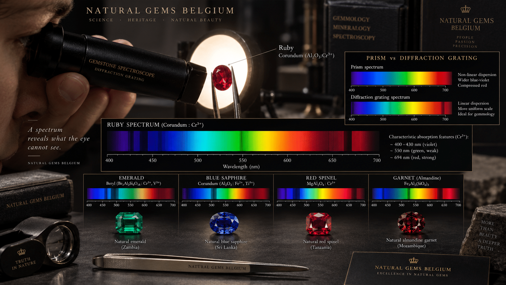
A Practical Guide to Absorption Lines, Bands and Gemstone Colour
A ruby appears red.
An emerald appears green.
A sapphire appears blue.
At first glance, colour seems to tell us what light a gemstone produces.
In most coloured gemstones, however, the opposite idea is more useful.
Ask:
Which parts of white light did the gemstone remove?
White visible light contains many wavelengths.
When light passes through a coloured gemstone, certain wavelengths can be absorbed more strongly than others.
The light that survives this selective absorption reaches our eyes.
That transmitted combination creates the colour we perceive.
A handheld spectroscope allows the gemmologist to separate transmitted light into its component colours and look directly for areas that have been absorbed.
Instead of seeing only:
red ruby
we can begin to see:
which wavelengths chromium absorbed to leave the transmitted red colour.
Instead of simply describing:
green emerald
we can investigate absorption associated with:
- chromium;
- vanadium;
- iron.
And in some gemstones, narrow absorption lines can provide highly useful supporting evidence for identification.
The spectroscope therefore adds another dimension to the practical gemmological sequence:
loupe → microscope → refractometer → polariscope → dichroscope → spectroscope.
The loupe and microscope reveal structure.
The refractometer measures refractive index.
The polariscope reveals optical anisotropy.
The dichroscope separates directional colours.
The gemstone spectroscope asks:
Which wavelengths of visible light are being absorbed?
Table of Contents
1. What Is an Absorption Spectrum?
Visible white light contains a continuous range of wavelengths.
For practical purposes, the human visible range extends approximately from:
violet and blue at shorter wavelengths
through:
green, yellow and orange
to:
red at longer wavelengths.
When white light is dispersed by a prism or diffraction grating, it appears as a coloured spectrum.
If a gemstone absorbs particular wavelengths, those regions become darker.
The resulting pattern is the gemstone’s:
absorption spectrum.
2. Why Gemstones Absorb Light
Gemstone colour arises from interactions between light and the material’s atomic and electronic structure.
Important mechanisms include:
- transition-metal ions;
- intervalence charge transfer;
- colour centres;
- structural defects.
Elements such as:
- chromium;
- vanadium;
- iron;
- titanium;
- manganese;
can contribute strongly to absorption in coloured gemstones.
The spectroscope allows some of those absorptions to be observed directly.
3. Absorbed Light Is Missing Light
This concept is fundamental.
Suppose a gemstone absorbs much of the:
- violet;
- blue;
- yellow-green
portion of the spectrum but transmits red strongly.
The stone can appear:
red.
Its colour is therefore determined not simply by what wavelengths are present, but by which wavelengths have been selectively removed from the incident light.
4. Transmission Creates Perceived Colour
The colour reaching the eye is the combined result of the wavelengths that remain after absorption.
GIA’s visible-spectroscopy research describes gemstone colour as the result of selective absorption of incident white light, with the unabsorbed portion transmitted to the observer.
This same principle applies from simple handheld spectroscope examination to sophisticated UV-Vis-NIR spectroscopy.
5. What Is a Handheld Gemstone Spectroscope?
A handheld gemmological spectroscope is a small optical instrument that disperses light into its component wavelengths.
The gemmologist examines light that has:
- passed through;
- or in certain situations reflected from
a gemstone.
Dark regions within the resulting spectrum indicate wavelengths where absorption is strong enough to be seen.
6. Two Main Spectroscope Designs
Two classical handheld designs are commonly used in gemmology:
Prism Spectroscope
Uses one or more prisms to disperse light.
Diffraction-Grating Spectroscope
Uses a diffraction grating to separate wavelengths.
Both reveal gemstone absorption spectra.
Their visual presentation differs.
7. Prism Spectroscope
A prism disperses different wavelengths by refraction.
Shorter and longer wavelengths are not spread uniformly.
Gem-A notes that its prism spectroscope produces a relatively wider blue region.
This can make spectral features in that part of the visible range easier to inspect.
8. Diffraction-Grating Spectroscope
A diffraction grating separates light through diffraction and interference.
Gem-A describes its diffraction spectroscope as producing a:
uniform, linear spectrum.
Wavelength intervals are therefore spread more evenly than in a prism instrument.
This can make the relationship between position and wavelength easier to visualise.
9. Neither Type Is Universally “Better”
Each design has practical strengths.
A prism instrument can provide greater apparent separation in some shorter-wavelength regions.
A diffraction instrument provides a more linear wavelength presentation.
Instrument familiarity often matters as much as theoretical preference.
The best instrument is one whose spectrum you can:
- focus;
- illuminate properly;
- read consistently.
10. GIA Uses a Diffraction-Grating Spectroscope
GIA’s current professional gem identification package includes a:
hand-held diffraction grating spectroscope.
Its current instrument is designed to produce a clear, bright spectrum using relatively low-intensity illumination.
This confirms that handheld visible spectroscopy remains part of modern classical gem identification.
11. Gem-A Uses Both Approaches
Gem-A currently offers:
- prism spectroscopes;
- diffraction-grating spectroscopes;
- focusing diffraction instruments.
Its portable gem instrument kit includes a diffraction-grating spectroscope.
The technique therefore remains relevant both in laboratory training and portable field testing.
12. What Does the Spectrum Look Like?
Without significant absorption, you would see a relatively continuous sequence:
violet → blue → green → yellow → orange → red
With a coloured gemstone in the light path, parts of this sequence may appear:
- darker;
- partially weakened;
- completely absorbed.
The dark features can take different forms.
13. Absorption Lines
A narrow dark feature is called an:
absorption line.
Some gemstones display very sharp lines associated with specific electronic transitions.
Narrow lines can be particularly useful because their approximate wavelength positions may be characteristic.
14. Absorption Bands
A broader dark region is called an:
absorption band.
Bands may cover a relatively large wavelength interval.
Their edges can be:
- sharp;
- gradual;
- diffuse.
Broad absorption is extremely important for gemstone colour.
But broad absorptions can be harder to interpret visually with a pocket spectroscope.
15. Cut-Offs
Sometimes one end of the spectrum becomes strongly absorbed over a wide region.
This can appear as a:
cut-off
where little useful transmitted light remains beyond a certain wavelength region.
Cut-offs can also contribute diagnostic information.
16. The Handheld Spectroscope Does Not Produce a Graph
This distinction is important.
With a pocket spectroscope, you visually see:
coloured light with dark absorption features.
A modern spectrophotometer instead records intensity numerically and produces a graph:
wavelength versus absorption or transmission.
That quantitative record is far more precise.
17. Handheld Versus UV-Vis-NIR Spectroscopy
A professional UV-Vis-NIR spectrometer can measure:
- absorption strength;
- exact wavelength;
- broad absorption envelopes;
- weak features;
- polarized spectra;
- wavelengths outside ordinary visible vision.
The handheld instrument cannot compete quantitatively.
Its advantage is:
- simplicity;
- speed;
- portability;
- immediate visual evidence.
18. Broad Absorption Can Be Hard to See
GIA’s research on coloured diamonds makes an important general point.
A handheld spectroscope can reveal sufficiently intense narrow absorptions as dark lines or bands.
But broad absorption regions can be much harder to recognise visually even though they may control much of the gemstone’s perceived colour.
This is one of the instrument’s central limitations.
19. Therefore “No Spectrum” Does Not Mean “No Absorption”
Every coloured gemstone must interact with visible light somehow.
If no diagnostic lines are visible in a handheld spectroscope, possible reasons include:
- absorptions are broad;
- absorptions are weak;
- the stone is too pale;
- the stone is too dark;
- the optical path is poor;
- illumination is inadequate;
- the instrument is not focused correctly.
Do not conclude that a gemstone has no absorption simply because you cannot see a useful pattern.
20. Prepare the Gemstone
Before testing, ensure the gemstone is reasonably clean.
Fingerprints and surface dirt can:
- scatter light;
- reduce transmission;
- make the spectrum harder to observe.
The stone does not need refractometer-style contact liquid.
This is a non-contact optical test.
21. Choose a Suitable Light Path
The objective is to send sufficient light through the gemstone and into the spectroscope.
For a faceted stone, experiment with different directions.
Useful paths may include:
- table to pavilion;
- pavilion to crown;
- through the girdle;
- through a thinner part of the stone.
The best path is usually the one providing a bright spectrum without overwhelming absorption.
22. Dark Stones May Need a Shorter Path
A strongly saturated gemstone may absorb so much light through its deepest direction that the entire spectrum becomes too dark.
Try:
- near the girdle;
- through a thinner region;
- through a lighter zone.
Reducing optical path length allows more light to reach the instrument.
23. Pale Stones May Need a Longer Path
A very pale gemstone presents the opposite problem.
Its absorption may be difficult to see because too little light is removed.
A longer path through the stone can strengthen visible absorption.
This relationship between:
path length and absorption
is a fundamental principle of spectroscopy.
24. Optical Path Length Matters
The farther light travels through an absorbing material, the more opportunity there is for absorption.
Therefore the same gemstone can show:
- stronger spectral features through a deep direction;
- weaker features through a shallow direction.
This is one reason repeat observations through different orientations can be useful.
25. Illumination Matters
A spectroscope can only analyse wavelengths actually supplied by the light source.
For classical gemmological work, use the illumination recommended for your specific instrument.
Some Gem-A spectroscope arrangements are specifically designed around incandescent illumination, while GIA’s current diffraction instrument can work with comparatively low-intensity light.
Do not assume every lamp produces an equally useful test spectrum.
26. First Observe the Light Source
Before placing the gemstone in the light path, look at your illumination through the spectroscope.
Become familiar with its baseline spectrum.
This helps distinguish:
features produced by the lamp
from:
features produced by the gemstone.
27. Focus the Spectroscope
Some spectroscopes have adjustable focus.
Gem-A’s focusing prism and diffraction models allow different spectral regions to be sharpened for the observer’s eyesight.
A poorly focused spectrum can turn:
- narrow lines;
- small bands
into vague dark areas.
Focus before interpreting.
28. Position the Slit Correctly
In instruments with an entrance slit, the transmitted gemstone light must enter that slit effectively.
If alignment is poor:
- the spectrum may be too dark;
- spectral features may disappear;
- stray light can dominate.
Small positional changes can make a large difference.
29. Shield Stray Light
Excess environmental light entering the eye or spectroscope reduces contrast.
For subtle spectra, shade the viewing area.
The goal is to see:
light transmitted through the gemstone
rather than reflections from the surrounding room.
30. Begin with Strong Training Stones
A beginner should not start with extremely weak spectra.
Useful teaching specimens include gemstones with reasonably recognisable absorption, such as:
- ruby;
- chromium-bearing spinel;
- some emeralds;
- certain garnets.
Building familiarity with strong examples makes later weak patterns easier to interpret.
31. Ruby: A Classic Spectroscope Stone
Ruby is one of the most important teaching examples.
Ruby is red corundum coloured primarily by:
Cr³⁺ — trivalent chromium.
GIA documents well-known chromium-related absorption in ruby, including broad absorptions in the violet/blue and green-yellow regions together with sharp chromium-related features in the red.
32. Why Ruby Appears Red
GIA’s polarized spectra of ruby show large Cr³⁺ absorption regions with transmission windows that leave significant red light available to the eye.
The balance of:
- absorbed wavelengths;
- transmitted red;
- sometimes chromium fluorescence
creates ruby’s characteristic appearance.
The handheld spectroscope lets the observer see part of this absorption architecture.
33. Chromium Features in Ruby
Quantitative GIA spectra document Cr³⁺ absorptions around:
- approximately 405–410 nm;
- a broad region around 560 nm;
together with the famous chromium feature near:
694 nm.
A handheld instrument may show only the stronger visually accessible portions depending on:
- stone;
- saturation;
- light source;
- optical path.
Do not expect a pocket spectroscope to reproduce a laboratory graph perfectly.
34. Fluorescence and Absorption Are Different
Ruby’s chromium can also produce strong:
red fluorescence.
Do not confuse this with absorption.
Absorption
Removes particular wavelengths from transmitted light.
Fluorescence
Emits new light after excitation.
The two phenomena can involve the same chromophore while representing different physical processes.
35. Blue Sapphire
Blue sapphire demonstrates a different colour mechanism.
Its blue colour commonly involves interaction between:
Fe²⁺ and Ti⁴⁺
in neighbouring sites within corundum.
This produces:
intervalence charge transfer.
GIA identifies a broad absorption centred approximately around 580 nm as a major contributor to blue corundum colour.
36. Why Blue Sapphire Can Be Harder in a Handheld Spectroscope
The Fe²⁺–Ti⁴⁺ absorption responsible for blue sapphire is broad.
Broad bands are inherently more difficult to characterise visually with a simple handheld spectroscope than narrow sharp lines.
This is a useful example of the instrument’s limitation.
A weak or indistinct handheld pattern does not mean sapphire lacks scientifically measurable absorption.
37. Corundum Can Have Multiple Chromophores
Natural corundum is rarely controlled by only one ideal chromophore.
GIA’s quantitative research identifies several important colour-producing systems in corundum, including:
- Cr³⁺;
- Fe³⁺;
- Fe²⁺–Ti⁴⁺;
- V³⁺;
- trapped-hole-related centres.
Natural spectra can therefore be combinations rather than textbook single-element patterns.
38. Real Gemstones Are Messier Than Diagrams
Teaching charts usually show simplified spectra.
A natural gemstone may contain several absorbers simultaneously.
Its observed spectrum can therefore contain:
- overlapping bands;
- weak lines;
- broad cut-offs;
- directional variation.
This is normal.
Do not reject a correct identification because a real specimen does not perfectly match an ideal classroom drawing.
39. Emerald
Emerald provides another classic spectroscopic system.
Its green colour is associated primarily with:
- chromium;
- vanadium;
with iron sometimes modifying the appearance.
Chromium- and vanadium-related absorptions can overlap substantially.
This is why detailed laboratory spectroscopy is often required for precise interpretation.
40. Chromium in Emerald
GIA emerald research documents Cr³⁺ absorption features approximately around:
- 430 nm;
- 600–630 nm;
with sharper chromium-related lines in the red region around:
- 637 nm;
- 646 nm;
- 658 nm;
- 680/683 nm
depending on composition and polarization.
Some of these sharp features can be useful in gemmological spectroscopy.
41. Vanadium-Dominant Emerald
Not every emerald is predominantly chromium-coloured.
Recent GIA research on emeralds from Zhen’an, China documents material in which:
V³⁺
is the dominant green chromophore.
Vanadium can produce broad visible absorption that overlaps chromium-related regions.
This makes the oversimplified statement:
“emerald spectrum = chromium”
scientifically unsafe.
42. Chromium and Vanadium Can Look Similar
GIA’s modern UV-Vis-NIR research notes that Cr³⁺ and V³⁺ can produce broadly similar absorption patterns in green gems.
Therefore a handheld spectroscope may support:
chromium/vanadium-type absorption
without necessarily allowing precise trace-element quantification.
Instrumental chemistry may be needed for exact interpretation.
43. Iron in Emerald
Iron can add further absorption features.
Depending on oxidation state and crystal site, iron-related absorption can occur in visible and near-infrared regions.
Some features may lie outside what an ordinary visible handheld spectroscope can evaluate comfortably.
Again, handheld observation is only one part of the analytical picture.
44. Red Spinel
Some red and pink spinels can display chromium-related absorption features.
GIA documented a natural dark pink-purple spinel in which a handheld spectroscope showed:
- thin bands near 680 nm;
- finer lines near 670 nm.
The spectrum contributed to the identification alongside:
- refractive index;
- specific gravity;
- fluorescence;
- microscopy.
This is exactly how the spectroscope should be used: as converging evidence.
45. Chromium Does Not Uniquely Mean Ruby
This is a critical interpretation rule.
Chromium can contribute colour to:
- ruby;
- emerald;
- spinel;
- garnet;
- tourmaline;
- diopside;
- other minerals.
Observing a chromium-type spectrum therefore does not identify the gemstone species by itself.
The host crystal still matters.
46. Chromium’s Pattern Depends on the Host Crystal
Cr³⁺ does not interact identically in every mineral.
The surrounding crystal field changes its electronic energy levels.
That is why chromium can contribute:
- red in ruby;
- green in emerald;
- green in chrome diopside;
- red or pink in spinel.
Spectroscopy reflects the interaction between:
chromophore + host structure.
47. Chrome Garnet
Chromium-bearing garnets can also display characteristic absorption.
But garnet chemistry is complex.
Different garnet members can contain combinations of:
- chromium;
- vanadium;
- iron;
- manganese.
Spectral interpretation should therefore be combined with:
- refractive index;
- specific gravity;
- colour;
- other tests.
48. Some Garnets Have Useful Iron Spectra
Iron-bearing garnets can show distinctive visible absorption patterns.
Historical and modern gemmological identification commonly uses combinations of bands rather than a single line.
A GIA case involving garnet, for example, documented handheld absorptions including features near:
- 505 nm;
- 527 nm;
- approximately 576 nm,
together with absorption toward the blue.
The complete gemmological dataset was still needed for final identification.
49. Spectra Can Support—but Not Replace—Other Tests
Suppose an unknown red stone displays chromium-type lines.
Possible candidates still include several species.
Now add:
- RI 1.762–1.770;
- DR uniaxial behaviour;
- ruby-type inclusions.
The evidence strongly converges toward:
ruby.
Spectroscopy becomes powerful when combined with independent properties.
50. The Spectroscope and Refractometer
These instruments answer very different questions.
Refractometer
Measures how strongly light changes velocity and direction in the gemstone.
Spectroscope
Shows where transmitted light is selectively absorbed.
A stone can share an RI range with another material yet display a different absorption spectrum.
The two tools therefore complement each other.
51. The Spectroscope and Dichroscope
Pleochroic gemstones absorb polarized light differently according to direction.
A dichroscope shows the resulting:
directional colours.
A spectroscope can reveal the:
directional absorption pattern
behind those colours.
Professional UV-Vis spectroscopy often records separate polarized spectra for different crystallographic vibration directions.
52. Ruby and Sapphire Spectra Can Be Polarized
Because corundum is anisotropic, absorption can differ according to optical direction.
GIA origin and colour research commonly records separate polarized spectra.
A handheld spectroscope usually provides a more qualitative observation.
But rotating a pleochroic stone can sometimes alter the apparent absorption pattern.
53. The Spectroscope and Microscope
Microscopy answers questions such as:
- natural or synthetic growth features?
- heat-altered inclusions?
- fissure filling?
- growth zoning?
The spectroscope answers:
- what visible wavelengths are being absorbed?
One cannot substitute for the other.
An emerald can have an emerald-like chromium spectrum yet still require microscopy to detect:
- doublet construction;
- filler;
- synthetic growth.
54. A Spectral Match Does Not Prove Natural Origin
Synthetic ruby is still chromium-bearing corundum.
Synthetic emerald can be chromium- or vanadium-bearing beryl.
Their absorption spectra may resemble those of natural equivalents.
Therefore:
A characteristic spectrum supports mineral/chromophore identification but does not automatically prove geological origin.
55. Treatment Detection Is Also Limited
Heat treatment can modify defects and chromophores.
In some cases, spectroscopy contributes important treatment evidence.
But the simple handheld instrument usually cannot determine reliably:
- heated versus unheated sapphire;
- diffusion treatment;
- subtle colour-centre modification.
Professional laboratories use quantitative spectroscopy and additional analytical techniques for those questions.
56. The Same Colour Can Have Different Spectra
Two gemstones may look nearly identical in colour while being coloured by completely different mechanisms.
A green stone might owe its colour mainly to:
- chromium;
- vanadium;
- iron;
- combinations of these.
A spectroscope can therefore provide information hidden behind visual colour.
57. Different Spectra Can Produce Similar Colours
GIA’s coloured-diamond spectroscopy research demonstrates a broad principle:
similar perceived colours can arise from different absorption patterns.
Colour alone therefore cannot reveal the underlying defect or chromophore reliably.
Spectroscopy helps expose those hidden differences.
58. Conversely, Similar Chromophores Can Produce Different Colours
Chromium is a perfect example.
The same oxidation state:
Cr³⁺
can contribute different colours depending on the host crystal structure.
Spectral interpretation must therefore always include the mineral context.
59. Dark Stones
A very dark gemstone may transmit too little light for a useful spectrum.
Try:
- a thinner area;
- edge illumination;
- stronger appropriate illumination;
- a different orientation.
If the entire visible spectrum remains nearly black, do not invent absorption lines.
Record:
spectrum not obtainable under test conditions.
60. Pale Stones
A pale gemstone may transmit almost all visible wavelengths strongly.
Diagnostic absorptions may be too weak for visual recognition.
Increasing optical path length can sometimes help.
But if no reliable feature appears, record that honestly.
61. Small Stones
Very small gemstones can be difficult because only a limited amount of transmitted light reaches the spectroscope slit.
Careful alignment becomes critical.
A stand or hands-free setup can make observation easier.
Gem-A specifically offers a larger teaching diffraction spectroscope with a rotating stand for easier demonstration and observation.
62. Mounted Gemstones
Mounted gems can often still be tested if light can be directed through them.
But metal settings may:
- block transmission;
- create reflections;
- limit orientation.
A spectroscope is often useful precisely because it can sometimes be applied without removing the stone from jewellery.
63. Opaque Materials
Classical transmitted-light handheld spectroscopy is most useful on:
- transparent;
- translucent
coloured materials.
Opaque stones may require other arrangements or more advanced reflected-light spectroscopy.
Do not assume the same simple technique applies equally to all gem materials.
64. Fluorescent Stones Can Complicate Observation
If the illumination strongly excites fluorescence, emitted light may contribute to what reaches the eye.
Absorption and emission are different processes.
For routine handheld work, interpret spectra conservatively when fluorescence is intense.
Advanced instruments separate these phenomena much more effectively.
65. Wavelength Numbers Versus Pattern Recognition
Some spectroscopes include wavelength scales.
Others encourage recognition of:
- approximate positions;
- pattern shapes.
Beginners often try to memorise dozens of exact nanometre values.
That is rarely the best first step.
First learn the spectrum’s structure:
- which end is red;
- which end is violet;
- where broad bands occur;
- where narrow lines cluster.
Then refine wavelength estimation.
66. Learn Colour-Wavelength Orientation
In a normal visible spectrum:
- violet/blue occupy shorter wavelengths;
- green lies in the middle;
- yellow/orange follow;
- red occupies longer wavelengths.
Approximate wavelength awareness helps translate a visual dark line into a meaningful gemmological observation.
67. Do Not Overstate Precision
If your pocket spectroscope shows a narrow line in the deep red but has limited scale precision, do not report:
681.37 nm
as though measured by a calibrated spectrophotometer.
Instead record something such as:
sharp absorption near 680 nm.
Measurement precision should match instrument capability.
68. Repeat Observations
Move the gemstone.
Change the light path.
Refocus.
Observe again.
A genuine absorption feature should be reasonably reproducible.
A dark patch caused by:
- poor alignment;
- stray light;
- facet obstruction
may disappear.
Repeatability improves confidence.
69. Compare Known Reference Stones
A powerful training method is to build a reference set.
Useful examples include:
- ruby;
- red spinel;
- emerald;
- chrome-bearing green gem;
- iron-bearing garnet;
- blue sapphire.
Observe their spectra repeatedly under the same conditions.
Eventually spectral patterns become visual memories rather than abstract textbook diagrams.
70. Record What You Actually See
A useful note might read:
Handheld spectrum: strong broad absorption in green-yellow region; sharp red lines visible near ~680–694 nm; violet region strongly absorbed.
That is far better than:
“Ruby spectrum.”
Description should precede interpretation.
71. Avoid Confirmation Bias
Suppose the stone looks like emerald.
Do not search desperately for an “emerald spectrum.”
Instead:
- observe;
- record;
- compare.
A spectrum inconsistent with your visual expectation is valuable evidence.
It may reveal:
- another gemstone;
- synthetic material;
- assembled material;
- unusual chemistry.
72. Broad Bands Are Easy to Misjudge
A gradually darkening spectral region can be harder to recognise than a sharp black line.
Our eyes are much better at detecting:
contrast boundaries
than subtle intensity gradients.
This is one reason quantitative spectrophotometry is superior for broad absorption.
73. Human Vision Is Part of the Instrument
In handheld spectroscopy, the detector is essentially:
your eye.
Different observers vary in:
- colour sensitivity;
- low-light vision;
- ability to recognise weak bands.
Practice improves reliability, but subjective limits remain.
Modern laboratory detectors remove much of this observer dependence.
74. Spectroscope Versus Spectrophotometer
A spectroscope lets you:
see the spectrum.
A spectrophotometer:
measures it.
The latter records numerical transmission or absorption intensities across wavelength.
This distinction explains why advanced laboratory spectroscopy can identify features that a handheld user cannot see.
75. UV-Vis-NIR Goes Beyond Human Vision
Professional UV-Vis-NIR instruments extend beyond ordinary visible wavelengths into:
- ultraviolet;
- near-infrared.
Important gemstone absorptions can occur outside the human visible range.
A handheld visible spectroscope therefore sees only part of the full optical spectrum.
76. Spectroscopy and Geographic Origin
Professional UV-Vis-NIR spectra can contribute to geographic-origin determination.
GIA uses spectral information alongside:
- microscopy;
- trace-element chemistry;
- reference collections.
For example, emeralds and sapphires from different geological environments can show different combinations of chromophore-related absorptions.
But the handheld spectroscope alone is not a country-of-origin instrument.
77. Emerald Origin Shows Why
Emerald populations can vary in relative:
- chromium;
- vanadium;
- iron
content.
Quantitative spectra can help characterise those differences.
Yet considerable overlap exists.
GIA therefore combines spectroscopy with trace-element chemistry and inclusion evidence rather than issuing geographic origin from one absorption line.
78. Sapphire Origin Is Similar
Sapphires from different deposits can differ in:
- Fe content;
- Fe-Ti absorption;
- other chromophore populations.
These spectral differences can support geological classification.
But professional geographic-origin work requires multiple independent data types.
A pocket spectroscope cannot say:
Sri Lanka
or:
Madagascar
with scientific reliability.
79. Spectroscopy and Treatment
Advanced spectroscopy can help investigate treatment because heating can modify:
- defects;
- oxidation states;
- chromophore relationships.
However, many treatment determinations require:
- FTIR;
- photoluminescence;
- UV-Vis-NIR;
- microscopy;
- trace-element analysis.
Do not overextend handheld visible spectroscopy.
80. Spectroscopy and Synthetic Identification
Laboratory-grown gems can possess different trace-element or defect environments from natural gems.
Advanced spectroscopy may exploit these differences.
But some synthetic material is deliberately grown to reproduce natural optical properties.
Therefore:
matching visible spectrum ≠ proof of natural origin.
81. Common Beginner Mistakes
Using the Wrong Light
The light source may not provide a useful continuous spectrum.
Looking Through Too Much Stone
Dark material absorbs nearly everything.
Looking Through Too Little Stone
Pale material may show weak absorptions.
Poor Alignment
Too little gemstone light enters the slit.
Not Focusing
Lines become fuzzy or disappear.
Memorising Numbers Without Patterns
Spectra become harder rather than easier to recognise.
Expecting Every Stone to Give a Diagnostic Spectrum
Many do not.
Treating a Chromium Spectrum as a Species Identification
Chromium occurs in many gems.
Confusing Absorption with Fluorescence
They are different physical phenomena.
Claiming Treatment or Origin from a Pocket Spectrum Alone
Usually unjustified.
82. Common Interpretation Errors
“The stone has chromium lines, so it is ruby.”
Incorrect.
Emerald, spinel, garnet and other minerals can contain chromium.
“I see no lines, so the stone has no chromophore.”
Incorrect.
Absorption may be broad or too weak for visual detection.
“The spectrum matches emerald, therefore it is Colombian.”
Incorrect.
Geographic origin requires much more evidence.
“Synthetic ruby will have a different basic chromium spectrum.”
Not necessarily.
Synthetic ruby is still chromium-bearing corundum.
“A dark region is automatically an absorption line.”
Not necessarily.
Check focus, alignment and reproducibility.
83. Evidence Classification
| Statement | Classification |
|---|---|
| Coloured gemstones selectively absorb portions of incident light | Established optical principle |
| A spectroscope disperses transmitted light into its component wavelengths | Established instrument principle |
| Dark lines and bands indicate relatively strong absorption regions | Established spectroscopic principle |
| Prism and diffraction-grating spectroscopes disperse light differently | Established optical principle |
| Diffraction instruments produce a comparatively linear wavelength spectrum | Established instrument behaviour |
| Chromium produces characteristic absorption in ruby | Established spectroscopic fact |
| Fe²⁺–Ti⁴⁺ intervalence charge transfer contributes to blue sapphire colour | Established corundum colour mechanism |
| Chromium and vanadium contribute important emerald absorption | Established spectroscopic fact |
| Broad absorption can be difficult to recognise visually with a handheld spectroscope | Established practical limitation |
| One absorption line uniquely identifies a gemstone | Incorrect |
| A handheld spectrum proves natural origin | Incorrect |
| A handheld spectrum normally determines geographic origin | Incorrect |
| No visible diagnostic spectrum means no absorption is present | Incorrect |
| Quantitative UV-Vis-NIR spectroscopy provides much more information than pocket visual spectroscopy | Established analytical fact |
84. A Repeatable Spectroscope Routine
Use the same sequence for every suitable gemstone.
Step 1 — Clean the Stone
Remove surface contamination.
Step 2 — Prepare the Light
Use an appropriate light source for the instrument.
Step 3 — Check the Baseline Spectrum
Observe the light source without the gemstone.
Step 4 — Position the Stone
Direct transmitted gemstone light into the spectroscope.
Step 5 — Focus
Sharpen the visible spectrum where the instrument permits.
Step 6 — Adjust Optical Path
Use a thinner or thicker part of the gemstone as needed.
Step 7 — Shield Stray Light
Improve contrast.
Step 8 — Scan the Whole Spectrum
Do not stare only at one expected wavelength.
Step 9 — Record Lines, Bands and Cut-Offs
Describe position and strength conservatively.
Step 10 — Rotate the Stone
Check whether orientation changes the spectrum.
Step 11 — Repeat
Confirm that features are reproducible.
Step 12 — Compare with Other Tests
Combine with:
- RI;
- polariscope;
- dichroscope;
- microscopy;
- fluorescence;
- specific gravity.
Step 13 — Escalate When Needed
Use laboratory spectroscopy when the question involves:
- treatment;
- synthetic origin;
- geographic origin;
- weak or overlapping chromophores.
85. A Practical Ruby Workflow
Unknown transparent red gemstone:
Refractometer
RI consistent with corundum.
Polariscope
Doubly refractive, uniaxial.
Dichroscope
Red/purplish-red directional colours.
Spectroscope
Chromium-related absorption.
Microscope
Natural corundum inclusion scene.
All of these independent observations converge toward:
natural ruby.
Treatment and geographic origin still require additional assessment.
86. A Practical Red Spinel Workflow
Unknown red gemstone:
Refractometer
RI around spinel range.
Polariscope
Singly refractive.
Dichroscope
No true pleochroism.
Spectroscope
Chromium-related red absorption features may be present.
The presence of chromium does not contradict spinel.
Instead, the entire dataset identifies the host mineral correctly.
87. A Practical Emerald Workflow
Unknown saturated green gemstone:
Refractometer
RI consistent with beryl.
Polariscope
Doubly refractive, uniaxial.
Dichroscope
Directional green/bluish-green colour.
Spectroscope
Chromium/vanadium-type absorption.
Microscope
Natural emerald inclusion scene.
The result strongly supports emerald.
The spectroscope still cannot establish:
- Colombia;
- Zambia;
- Afghanistan;
- oil versus resin;
- natural versus hydrothermal synthetic on its own.
88. Why Combining Instruments Works
Each instrument interrogates a different physical property.
Loupe / Microscope
What structures are visible?
Refractometer
How strongly is light refracted?
Polariscope
Does optical behaviour change with direction?
Dichroscope
Does colour absorption change with direction?
Spectroscope
Which wavelengths are absorbed?
When several independent properties agree, identification confidence rises dramatically.
89. The Spectroscope Teaches a Larger Lesson About Colour
Gemstone colour is not simply a label:
red, blue, green.
It is the visible result of specific atomic-scale interactions with light.
A ruby is red because certain wavelengths are strongly absorbed while others are transmitted.
An emerald is green because its chromium and/or vanadium environment produces a different absorption pattern.
Blue sapphire owes its colour to another mechanism again.
The spectroscope makes those invisible atomic interactions visible.
Conclusion
A handheld spectroscope is one of the smallest instruments in a gemmological kit.
Yet it opens a direct window into the physics of gemstone colour.
White light enters the gemstone.
Some wavelengths pass.
Others are absorbed.
The spectroscope separates what survives into a visible spectrum.
Dark lines reveal narrow absorption.
Broad dark regions reveal wider absorption bands.
Cut-offs show where transmission becomes strongly reduced.
In ruby, chromium leaves characteristic spectral evidence.
In emerald, chromium and vanadium create overlapping green-gem absorption systems.
In sapphire, iron and titanium can produce broad absorption associated with blue colour.
In spinel, garnet and many other species, additional chromophores produce their own patterns.
But the instrument must be used with restraint.
A chromium spectrum does not automatically mean ruby.
A green spectrum does not identify geographic origin.
A matching natural-gem spectrum does not automatically prove that a specimen is natural rather than laboratory-grown.
A lack of visible lines does not mean that no absorption exists.
And broad absorption that dominates gemstone colour may be difficult to recognise with the human eye through a pocket instrument.
That is why the professional workflow remains:
observe → record → compare → combine → conclude.
Use the spectroscope beside the:
- refractometer;
- polariscope;
- dichroscope;
- microscope.
Let each instrument contribute independent evidence.
And when the question becomes more sophisticated—treatment, synthetic origin, trace-element chemistry or geographic origin—move from visual spectroscopy to quantitative laboratory methods such as:
UV-Vis-NIR spectroscopy.
The handheld spectroscope does not replace modern analytical instrumentation.
It teaches the principle on which much of that instrumentation is built.
It transforms gemstone colour from something we merely admire into something we can begin to understand scientifically.
References & Further Reading
Gemological Institute of America — Diffraction Grating Spectroscope.
Current GIA professional instrument reference. GIA describes its compact handheld diffraction-grating instrument as producing a clear, bright visible spectrum with relatively low-intensity illumination.
GIA Diffraction Grating Spectroscope
Gemological Institute of America — Gem Identification Student Package.
Current professional training reference confirming that a handheld diffraction-grating spectroscope remains part of GIA’s standard gem-identification equipment alongside the refractometer, polariscope and dichroscope.
GIA Gem Identification Student Package
Gem-A — Diffraction Grating Spectroscope.
Current professional instrument reference describing the use of a diffraction grating for coloured gem materials and its production of a relatively uniform linear visible spectrum.
Gem-A Diffraction Grating Spectroscope
Gem-A — Prism Spectroscope.
Current Gem-A reference describing prism dispersion and the relatively greater spread of the blue region that can help observation of absorption features at shorter visible wavelengths.
Gem-A Prism Spectroscope
Gem-A — Focussing Diffraction Grating Spectroscope.
Professional reference for an adjustable-focus handheld instrument that allows spectral regions to be sharpened for the user’s eyesight.
Gem-A Focussing Diffraction Grating Spectroscope
Gem-A — Portable Gem Instrument Kit.
Current evidence for the continuing practical role of handheld spectroscopy in field gemmology. Gem-A includes a diffraction-grating spectroscope alongside the loupe, polariscope, conoscope and dichroscope in its portable professional kit.
Gem-A Portable Gem Instrument Kit
Shigley, J. E. & Breeding, C. M. — “Visible Absorption Spectra of Colored Diamonds.” Gems & Gemology, Spring 2015.
Important explanation of visual versus instrumental spectroscopy. It shows that a handheld spectroscope can reveal intense narrow absorptions, while broad absorption regions may be difficult to observe despite their major contribution to gemstone colour. GIA — Visible Absorption Spectra of Colored Diamonds
Breeding, C. M. et al. — “Application of UV-Vis-NIR Spectroscopy to Gemology.” Gems & Gemology, Winter 2024.
Current comprehensive review of quantitative optical spectroscopy in gemmology, including chromophore identification, chromium/vanadium absorption, fancy-colour diamonds and the use of UV-Vis-NIR spectra in advanced laboratory analysis. GIA — Application of UV-Vis-NIR Spectroscopy to Gemology
Dubinsky, E. V., Stone-Sundberg, J. & Emmett, J. L. — “A Quantitative Description of the Causes of Color in Corundum.” Gems & Gemology, Spring 2020.
Major scientific reference for corundum chromophores, including Cr³⁺, Fe³⁺, Fe²⁺–Ti⁴⁺, V³⁺ and trapped-hole-related colour centres and their quantitative absorption behaviour. GIA — Causes of Color in Corundum
Groat, L. A. et al. — “A Review of Analytical Methods Used in Geographic Origin Determination of Gemstones.” Gems & Gemology, Winter 2019.
Authoritative overview demonstrating how ruby and sapphire absorption spectra contribute to modern origin research alongside microscopy and trace-element chemistry. Includes Cr³⁺ absorption in ruby and Fe²⁺–Ti⁴⁺ intervalence charge-transfer absorption in blue sapphire. GIA — Analytical Methods for Geographic Origin Determination
Guo, Y., Zheng, Y.-Y. & Yu, X.-Y. — “Characterization of Emeralds from Zhen’an County in Shaanxi, China.” Gems & Gemology, Winter 2025.
Recent emerald study demonstrating a vanadium-dominant colour mechanism and illustrating why green beryl/emerald absorption cannot simply be interpreted as chromium alone. GIA — Emeralds from Zhen’an, China
Saeseaw, S., Pardieu, V. & Sangsawong, S. — “Three-Phase Inclusions in Emerald and Their Impact on Origin Determination.” Gems & Gemology, Summer 2014.
Useful emerald spectroscopy reference documenting absorption associated with Cr³⁺, V³⁺ and Fe in emerald and showing how spectroscopy is integrated with microscopic and chemical evidence. GIA — Three-Phase Inclusions and Emerald Origin
Cooper, A. & Sun, Z. — “Spinel Inclusion in Spinel.” Gems & Gemology, Winter 2014.
Practical laboratory example in which a handheld spectroscope revealed red-region absorption features near 680 nm and 670 nm in a natural pink-purple spinel, while RI, SG, fluorescence and microscopy completed the identification. GIA — Spinel Inclusion in Spinel
Gemological Institute of America — “Emerald/Emerald Doublet.” Gems & Gemology, Spring 2016.
Excellent reminder of spectroscope limitations: a chromium spectrum consistent with emerald was present, but microscopy was required to reveal that the specimen was an assembled emerald doublet. GIA — Emerald/Emerald Doublet